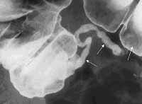
Normal appendix. Complete contrast-filled appendix is observed, which effectively excludes the diagnosis of appendicitis on barium enema examination (arrows).
Anatomy: Appendix vermiformis is a thin, wormlike, tubular organ that is located at the inferior part of the cecum. In adults, a normal vermiform appendix varies in length from 5-35 cm (average 8 cm).
The appendix has no fixed position. It originates 1.7-2.5 cm below the terminal ileum, either in a dorsomedial location (most common) from the cecal fundus, directly beside the ileal orifice, or as a funnel-shaped opening (in 2-3% of patients). The appendix has a retroperitoneal location in 65% of patients and may descend into the iliac fossa in 31%. The difference in appendiceal position changes clinical findings considerably.
Appendiceal congenital disorders are extremely rare but occasionally reported (eg, agenesis, duplication, triplication).
The appendix has its own mesentery arising from a peritoneal extension extending from the terminal ileum to the medial aspect of the cecum and appendix. This fold contains the appendicular artery, which is a terminal branch of the ileocolic artery, and runs adjacent to the appendicular wall. Venous drainage is via the ileocolic veins and the right colic vein into the portal vein, and lymphatic drainage occurs to the ileocolic nodes along the course of the superior mesenteric artery to the celiac nodes and cisterna chyli.
The appendix is contained within the visceral peritoneum forming the serosa and its exterior layer is longitudinal muscle with a circular muscle layer deep to the longitudinal muscle. Beneath this lies the submucosal layer containing lymphoepithelial tissue. The mucosa consists of columnar epithelium with few glandular elements and neuroendocrine argentaffin cells.
But in this case it's actually second part of Local Distortion remix.